Blog
If you’ve never had Mohs surgery (the surgical technique to treat skin cancers) it can sound confusing, or even a little overwhelming. Many people are unaware of this procedure until either themselves, or a loved one needs to have it done. In this week’s blog, you’ll learn about the evolution of skin cancers, the differences between them, and breakdown the Mohs surgery procedure to help you feel more at ease and understanding of what to expect.
While normal skin cells grow, develop, and die in predictable cycles, skin cancer develops when skin cells grow out of control. Instead of dying, the damaged DNA within skin cancer cells causes them to continue growing and produce more abnormal cells. They also then can invade other tissues. Skin cancer is the most common type of cancer in the US.
The primary cause of skin cancer is exposure to ultraviolet (UV) light which damages the DNA with repeated exposure. People with chronic exposure to UV light, whether in the outdoors or in tanning beds, are at increased risk of developing skin cancer. Recently the World Health Organization elevated tanning beds to its highest cancer risk category, the same rating it gives to cigarettes. Immunosuppressed patients, such as organ transplant recipients or patients with chronic lymphocytic leukemia are at a greatly increased risk as well, because their immune systems are not as capable of warding off cancerous cells.
The skin is made up of several types of cells which can be affected by distinct types of skin cancer. The three most common types of skin cancer, which together make up approximately 99% of diagnosed cases, are basal cell carcinoma, squamous cell carcinoma, and melanoma.
As the most common type of skin cancer, basal cell carcinoma accounts for about 80% of all diagnosed skin cancers. It begins in the keratinocyte cell of the epidermis, which is located in the lowest layer of the epidermis. This type of cancer can look like a sore that doesn’t completely heal, a shiny bump, or a reddish, irritated area usually in sun exposed skin. It usually progresses slowly and does not metastasize or spread to distant parts of the body. Eventually, though if untreated it can invade and damage local muscle, nerves, and even cartilage or bone.
Potentially more aggressive than basal cell carcinoma, squamous cell carcinoma forms just beneath the surface of the skin in the keratinocyte cell of the epidermis. While this second most common type of skin cancer usually develops on sun-exposed skin, it can form on other areas of the body like mucous membranes and genitalia. It often looks like a thick, rough, scaly patch or wart that can grow rapidly. While the vast majority of squamous cell carcinomas do NOT spread or metastasize to distant areas, there is a potential risk of this particularly when it arises on the lip or ear, or in an immunocompromised patient.
The most dangerous of the common forms of skin cancer is melanoma. While it accounts for only about 3% of total skin cancer cases, melanoma is responsible for over 75% of skin cancer related deaths. Nearly 10,000 Americans die of melanoma each year. Melanoma originates in the pigment-producing cells called melanocytes, which give the hair, skin, and eyes their color. Melanomas are usually black or dark brown and can develop in a new or preexisting mole. If identified early when the melanoma is still very thin, the melanoma can be removed with local anesthesia in the office. Once the melanoma has grown deeper, then it must be removed as well as lymph nodes in the area, which requires surgery to be done in an operating room under general anesthesia.
There are many acceptable skin cancer treatment options aside from Mohs surgery such as freezing or cryosurgery, scraping and burning, topical chemo creams, surgical excision, radiation and laser surgery. There are advantages to Mohs surgery compared to other skin cancer treatments though:
- It offers the highest cure rate of all treatment options for all skin cancers but especially if the area is recurrent from a previous treatment or located in scar tissue
- It preserves the most healthy/unaffected surrounding skin so it is especially appropriate on preserving cosmetic appearance and function on the head and neck area. Additionally, it is useful in areas where the skin is tight or lacking laxity such as the scalp, lower leg, hands, and feet.
- Larger skin cancers (greater than 2 cm), cancers with ill-defined edges, and cancers that are growing rapidly are also optimally treated with Mohs micrographic surgery.
The term “Mohs” refers to Dr. Frederic Mohs, a surgeon at the University of Wisconsin who developed the surgical technique in the 1930’s. The technique has undergone many refinements but has come to be known as Mohs surgery in honor of Dr. Mohs. Dr. Mohs recognized that a skin cancer can resemble the “tip of the iceberg” with more tumor cells growing under the skin similar to roots of a tree. Patients often come to their surgery appointment after they have healed from their biopsy and question why they need to have anything done since the area looks better than it did before the biopsy. I use the tree analogy stating that the biopsy cut down the tree so from a distance the tree appears to be gone but now the stump and/or roots need to removed or the tree will grow back.
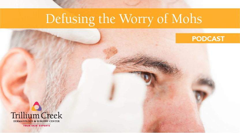
So what can one expect on the day of surgery: Initially I will come in and exam and mark the area to be removed. Using a scalpel, a very thin rim usually 1-2 millimeters is removed around and taken to the lab for processing. Any bleeding then is stopped with a combination of holding pressure and an electrocautery tool. A temporary bandage is placed on the site and the patient is then allowed to wait in one of our waiting areas while the tissue is processed. This initial procedure usually takes about 5-10 minutes. Depending on the size of the initial piece, the processing time can vary from 30-90 minutes (45 minutes is typical though).
Once the tissue is removed, it is brought into our lab where the edges are marked with various colored dyes for orientation purposes, and a map of the specimen is created. The tissue is then frozen solid so that it can be sectioned into very thin slices (1/20 the thickness of a sheet of paper). These slices are then placed on several glass slides which are then sent through an automated staining device. Once the slides come out of the staining device, I can then examine them under the microscope allowing me to check all of the deep and peripheral margins of the piece of tissue. If cancer cells are identified at any margin, I then mark on the map where this is in relation to the dyes that were placed prior to processing. The patient is then brought back into the exam room. More anesthesia is administered (however this is done before the initial has worn off so it resets the clock on when the numbness will wear off) and an additional tissue layer is removed ONLY in the area or areas where the cancer is still present, leaving normal skin intact.
Once the complete removal has been confirmed, I will then explain the options for repair of the wound. Usually this involves suturing or stitching the wound closed although sometimes letting the wound heal on its own is a viable option. Occasionally, a wound will need a skin graft or a skin flap (where local skin is loosened and pulled into an adjacent area). This is discussed with the patient before any repair is begun. In my practice we have found that about 80% of the time the tumor will be removed with the first piece and only 5% of tumors will require three or more layers to adequately remove the tumor cells. Most patients end up spending 1-2 hours at our office on the day of their Mohs surgery but 3-4 hours is possible for the 5% that require three or more layers. After the wound has been repaired, a pressure bandage is placed over the site and typically left in place for 1-2 days. We ask that the patients avoid any strenuous activity for 24-48 hours to avoid bleeding and swelling complications and usually suggest applying ice to the area intermittently for the rest of the day to avoid swelling and ultimately bruising around the site. Generally, postoperatively patients can easily manage their soreness with over the counter medications such as ibuprofen or acetaminophen for the first 1-2 days, rarely needing anything after that. Patients typically continue their normal medications before and after the surgery with the exception of those individuals who take an aspirin purely for preventative reasons. Those patients who take a prescription blood thinner will typically continue them, realizing that they often lead to significant bruising which can take 7-10 days to resolve.
After 1-2 days the patient is asked to remove the initial bandage and then wash the area gently with soap and water. If any dried blood or crust has developed that does not come off with gentle cleansing, we ask the patient to apply hydrogen peroxide on q-tips to the area to gently lift off the scab or crust. Then apply Vaseline or Aquaphor to the site and cover with a non stick pad or Band-Aid. This is done daily until the sutures are removed usually in 7-10 days. Typically, once the sutures are removed, bandaging the site is no longer needed. Depending on the location it will usually take 1-3 months for any redness to completely dissipate and for the scar to soften and become inconspicuous. I recommend using silicone scar gels and daily massage (using a lotion or cream to minimize friction) to expedite the scar fading process. The silicone gels can usually be started within 1-2 days of removing the suture and the massage can be initiated 7-10 days after suture removal.
If you would like additional information, visit TCOhio.com. To schedule an appointment, call us at 330.725.0569.